Atrial Septal Defect Closure
An atrial septal defect (ASD) is one of the most common congenital heart conditions. It is an opening in the wall that separates the two upper chambers of the heart, the right and left atria. The presence of this opening allows blood to pass from one atrium to the other, affecting normal blood circulation.
Although in some cases an atrial septal defect may be small and not cause significant problems, larger defects require medical treatment. Closure of an atrial septal defect is an effective therapeutic option that restores normal heart function and reduces the risk of serious complications.
What is an Atrial Septal Defect
The heart is divided into four chambers: two atria and two ventricles. The atrial septum is the wall that separates the right atrium from the left atrium.
In an atrial septal defect, there is an opening in this septum, allowing blood to flow from the left atrium to the right atrium. This results in increased blood flow to the lungs and puts strain on the right side of the heart.
The condition is usually congenital, meaning it is present from birth, but it is often diagnosed later in childhood or even in adulthood.
What is the Mediastinum
The mediastinum is the central space of the chest, located between the two lungs. In this area, important structures are found, such as:
- the heart
- the trachea
- the esophagus
- major blood vessels
- lymph nodes
- the pericardium
Tumors that develop in this area may cause pressure on vital organs and lead to serious symptoms.
Most Common Types of Tumors
Most common tumor types
Tumors that appear in the heart and mediastinum include:
Benign heart tumors
The most common is the cardiac myxoma, a benign tumor that usually appears in the left atrium.
Other benign tumors include:
- fibromas
- lipomas
- rhabdomyomas
When is Closure of the Defect Needed
The decision for closure is made after a cardiological evaluation, which includes tests such as:
- echocardiogram
- transesophageal echocardiography
- cardiac catheterization
- cardiac MRI
Closure is recommended when:
- the defect is large
- it causes symptoms
- there is significant strain on the heart
- there is an increased risk of complications
Early treatment can prevent long-term damage to the cardiovascular system.
Methods of Atrial Septal Defect Closure
Modern medicine offers two main treatment methods:

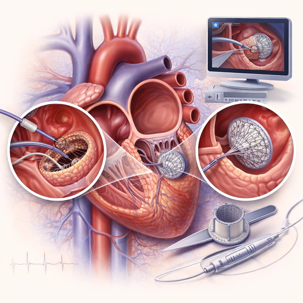
Transcatheter Closure
In some cases, the defect can be closed using a catheter. The procedure is performed through the femoral vein, and a special device is placed to seal the opening.

Surgical Closure
When the defect is large or cannot be treated with a catheter, surgical closure is performed. During the procedure, the cardiac surgeon closes the opening either with sutures or with a special biological material. The surgical method provides a definitive solution with very high success rates.
Modern Cardiac Surgery Techniques
Advances in cardiac surgery have enabled the application of more modern techniques, such as endoscopic cardiac surgery, which is performed through smaller incisions.
The advantages of this technique include:
- smaller surgical trauma
- less postoperative pain
- faster recovery
- shorter hospital stay
- better aesthetic outcome
The choice of the appropriate technique depends on the characteristics of the condition and the patient’s overall health.
Aortic Valve Replacement
When the valve cannot be repaired, replacement is performed. The damaged valve is removed and replaced with an artificial valve.
The two main types of valves are:
Mechanical Valves
They are highly durable and have a long lifespan, but require lifelong anticoagulant therapy.
Biological Valves
They are made from biological tissues and usually do not require long-term anticoagulant therapy, but they have a shorter lifespan.
The choice of the appropriate valve depends on the patient’s age, medical history, and other clinical factors.
(FAQ)
It is an opening in the wall that separates the two atria of the heart.
If left untreated, it can lead to pulmonary hypertension, cardiac arrhythmias, and heart failure.
The procedure is recommended when the defect is large or causes symptoms.
Surgical closure usually lasts 2 to 4 hours.
Full recovery may take from a few weeks to a few months.
Yes. After treatment, most patients fully return to their daily activities.
With significant experience in international medical centers and numerous scientific distinctions, cardiac surgeon in Athens Dr. E. Andreas Agathos is a reliable choice for patients seeking specialized cardiac surgical care.
He is a Doctorate holder from the University of Athens and holds important international titles:
- FRCS – Royal College of Surgeons of Edinburgh
- FACS – American College of Surgeons
- FETCS – European College of Surgeons
- FICS – International College of Surgeons
Dr. Agathos also holds multiple international patents (World patent, USA patent, European patent) for the development of new biological materials, innovative heart valves, and anti-calcification treatment methods.
He has served as Director of Cardiac Surgery at hospitals such as Hygeia, Athens Medical Center, and Euroclinic Athens, while also collaborating with leading international hospitals such as Massachusetts General Hospital in Boston and Triemli Hospital in Zurich.
With more than 5,000 surgeries in Greece and abroad, he has particular expertise in endoscopic cardiac surgery, offering modern and less invasive solutions for patients.
His international experience, scientific innovation, and globally recognized patents in the field of cardiac valves make Dr. Agathos one of the most specialized cardiac surgeons in modern cardiac surgery.

